|
|
Medical image quality enhancement |
|
|
Medical image quality enhancement |
|
|
|
|
|
|
|
|
|
|
|
|
|
|
|
|
|
|
|
|
|
|
Dual-energy computer tomography
The research is supported by the Economic Developmental Operative Program with the cooperation of General Electric and the University of Szeged.
Introduction:
The electrons, accelerated by corresponding electric field, can produce such high energy photons, that pass through organic and inorganic materials. The spectrum of the generated X-ray radiation characterizes the material of the anode.
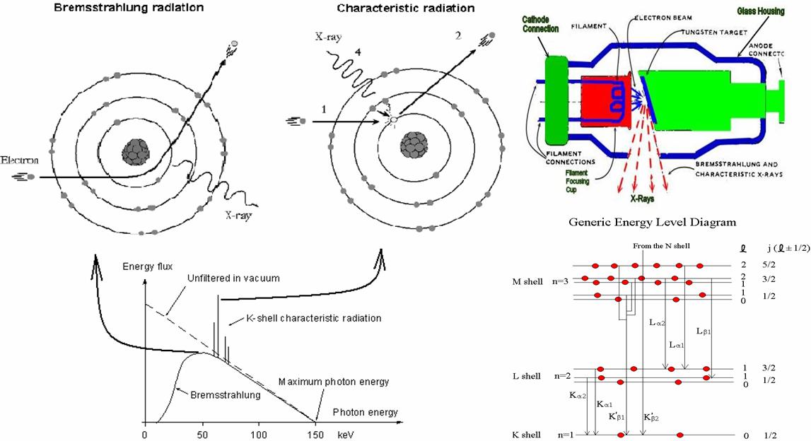
The extent of the absorption depends on the photon energy and the compound of the absorbing material, hence the absorbed X-ray photons depict shadow image on the detector. Materials with similar absorption give similar shadow image, so they can be hardly distinguished. But the modern dual-energy equipments can differnetiate materials with an additional projection (using different energy). Materail specific images (bone image, soft tissue image) can be obtained from dual energy exposures. Such equiment is the dual energy CT.
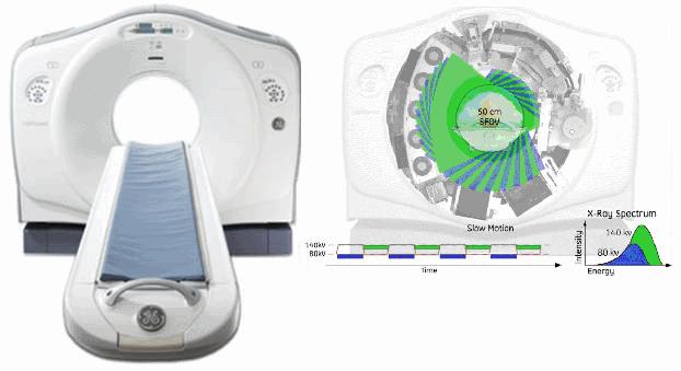
General Electric CT 750 HD
Objectives:
Practical application of the plus information comes from Dual-energy CT device (DECT), study the separability of the different tissues.
Duration: 1,5 years
Required skills:
Interests in medical imaging, programming knowledge, english, skills in mathematics ang algorithm development.
Partial tasks: