|
|
Medical image quality enhancement |
|
|
Medical image quality enhancement |
|
|
|
|
|
|
|
|
|
|
|
|
|
|
|
|
|
|
|
|
|
|
The team, formed to study and develop the medical imaging techniques, started working in 2003 with the support of the GE Healthcare. The basic goal of the first project (March 2003 - June 2003: Beam Hardening and Metallic Artifacts Correction Techniques in Computerized Tomography) was to understand analyse the correction techniques for image degrading effects in CT imaging known as beam hardening and metallic artifacts. The project came true by cooperating with the Department of Image Processing and Computer Graphics. More than ten students participated in the literature review part. 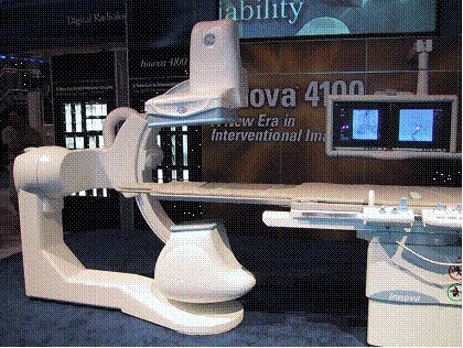
As the continuation of this successful project, another three months long project (XTOP corretcion) started in June 2004, which aimed to analyse and make proposals to correct the physical modells of the XTOP simulator tool developed by GE to improve precision of simulations. During the project, spectra of X-ray sources, used in tomography and the scatterig processes of different tissues were studied in detail. Besides, experimental measurements were also performed with Innova 4100 in the center of GEMS, in Teve street.