Localization microscopy method
The spatial resolution of a traditional optical imaging 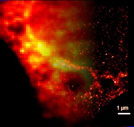 system is limited by its point-spread function (PSF). Point-like sources with a spatial separation smaller than the width of the PSF cannot be separated and will be detected indistinguishably. However, in the localization method the activation of the point-like sources (fluorescent proteins, dyes, etc.) is separated in time and can be detected independently. The centre of the PSF can be determined by fitting, and hence the exact position of the source can be localized with a precision of <20nm. Super-resolution images can be generated by sequentially repeating the activation-localization steps.
Recommended publications:
- Lelek, M., Gyparaki, M.T., Beliu, G. et al. Single-molecule localization microscopy. Nat Rev Methods Primers 1, 39 (2021). doi:10.1038/s43586-021-00038-x
- van de Linde, S., Löschberger, A., Klein, T., Heidbreder, M., Wolter, S., Heilemann, M., & Sauer, M. (2011). Direct stochastic optical reconstruction microscopy with standard fluorescent probes. Nature protocols, 6(7), 991-1009
doi:10.1038/nprot.2011.336 - Dempsey, G., Vaughan, J., Chen, K. et al. Evaluation of fluorophores for optimal performance in localization-based super-resolution imaging. Nat Methods 8, 1027–1036 (2011).
doi:10.1038/nmeth.1768 - Heilemann, M., Van De Linde, S., Schüttpelz, M., Kasper, R., Seefeldt, B., Mukherjee, A., … & Sauer, M. (2008). Subdiffraction‐resolution fluorescence imaging with conventional fluorescent probes. Angewandte Chemie International Edition, 47(33), 6172-6176.
doi:10.1002/anie.200802376 - Betzig, E., Patterson, G. H., Sougrat, R., Lindwasser, O. W., Olenych, S., Bonifacino, J. S., … & Hess, H. F. (2006). Imaging intracellular fluorescent proteins at nanometer resolution. Science, 313(5793), 1642-1645.
doi:10.1126/science.1127344 - Erdélyi Miklós, Sinkó József: Optikai pointillizmus: A lokalizációs optikai mikroszkópia, Fizikai Szemle 2014.05.
Optical beam engineering
Optical imaging can be enhanced by either modifying the 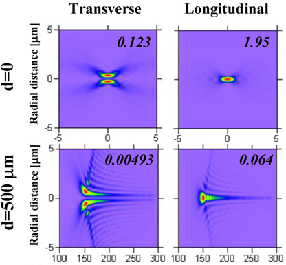 illumination or the detection beams. Wide-field structured illumination excites the sample with a sinusoidal pattern and can improve the spatial resolution by a factor of two. In the scanning methods (confocal microscopy, etc.) the PSF (either its shape or polarization) can be modified to improve the spatial resolution and depth of focus. Radially and azimuthally polarized beams can be generated by means of a birefringent plate. But PSF engineering can be also applied in the detection path for 3D super-resolution imaging.
Tomographic Optical Microscopy
Tomography, (e.g.: CT, PET, SPECT, μCT, etc.) is one of the most widely applied methods in medical imaging. This method can be adapted to the optical regime, and various tomographic imaging techniques have been developed such as optical projection tomography, optical coherence tomography, tomographic diffractive microscopy (TDM), and LSTOM. Because tomography is a polar data acquisition  technique, it requires a precise rotating system where the center of rotation (COR) must be known with high accuracy. To avoid COR error, geometrical calibration and post-processing algorithms are used to realign the projections and reconstruct the error-free images. However, when the voxel size decreases—for example in case of μCT and TDM—the accuracy of the rotation and alignments becomes more important. Furthermore, mechanical and thermal drifts also have a more critical effect on the imaging.

You must be logged in to post a comment.