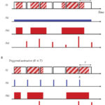
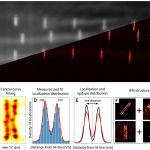
Machine learning framework to segment sarcomeric structures in SMLM data

Object detection is an image analysis task with a wide range of applications, which is difficult to accomplish with traditional programming. Recent breakthroughs in machine learning have made significant progress in this area. However, these algorithms are generally compatible with traditional pixelated images and cannot be directly applied for pointillist datasets generated by single molecule localization microscopy (SMLM) methods. Here, we have improved the averaging method developed for the analysis of SMLM images of sarcomere structures based on a machine learning object detection algorithm. The ordered structure of sarcomeres allows us to determine the location of the proteins more accurately by superimposing SMLM images of identically assembled proteins. However, the area segmentation process required for averaging can be extremely time-consuming and tedious. In this work, we have automated this process. The developed algorithm not only finds the regions of interest, but also classifies the localizations and identifies the true positive ones. For training, we used simulations to generate large amounts of labelled data. After tuning the neural network’s internal parameters, it could find the localizations associated with the structures we were looking for with high accuracy. We validated our results by comparing them with previous manual evaluations. It has also been proven that the simulations can generate data of sufficient quality for training. Our method is suitable for the identification of other types of structures in SMLM data.

https://doi.org/10.1038/s41598-023-28539-7

Quantitative dSTORM superresolution microscopy

Localization based superresolution technique provides the highest spatial resolution in optical microscopy. The final image is formed by the precise localization of individual fluorescent dyes, therefore the quantification of the collected data requires special protocols, algorithms and validation processes. The effects of labelling density and structured background on the final image quality were studied theoretically using the TestSTORM simulator. It was shown that system parameters affect the morphology of the final reconstructed image in different ways and the accuracy of the imaging can be determined. Although theoretical studies help in the optimization procedure, the quantification of experimental data raises additional issues, since the ground truth data is unknown. Localization precision, linker length, sample drift and labelling density are the major factors that make quantitative data analysis difficult. Two examples (geometrical evaluation of sarcomere structures and counting the γH2AX molecules in DNA damage induced repair foci) have been presented to demonstrate the efficiency of quantitative evaluation experimentally.

Novák, T., Varga, D., Bíró, P., H. Kovács, B. B., Majoros, H., Pankotai, T., … & Erdélyi, M. (2022). Quantitative dSTORM superresolution microscopy. Resolution and Discovery. https://doi.org/10.1556/2051.2022.00093

Analysis of Ionizing Radiation Induced DNA Damage by Superresolution dSTORM Microscopy

The quantitative detection of radiation caused DNA double-strand breaks (DSB) by immunostained γ-H2AX foci using direct stochastic optical reconstruction microscopy (dSTORM) provides a deeper insight into the DNA repair process at nanoscale in a time-dependent manner.

Read more

FOM2021 presentation

WE-PAR2-B Fluorescence V: Probes, labelling, Bio Applications II 
(Room B, 2021.03.31.)

  • 15:10-15:25: Anti-Stokes Fluorescence Microscopy with Alexa Fluor 568.
    T. Gajdos, M. Erdélyi (University of Szeged, Department of Optics and Quantumelectronics, Hungary)

Full Program >>

[HU] Introduction video for students

Lab intriduction video for students (2021):

…and how we moved in in 2014:

Read more

PhD graduation

Our college, Tamás Gajdos has successfully completed the Physics PhD program at the University of Szeged. His dissertation was written under the title „Superresolution localization microscopy using multiple modalities” and was defended with a result of 96% on November 20th. The dissertation and the thesis synopsis can be accessed from SZTE Repository of Dissertations.

Read more

Syndecan-4 Modulates Cell Polarity and Migration by Influencing Centrosome Positioning and Intracellular Calcium Distribution

In collaboration with the Muscle Adaptation Group, we have published a new paper in Frontiers in Cell and Developmental Biology. Our join research venture studied the regeneration process in the tissue on a molecular level.

Becsky, Daniel, et al. “Syndecan-4 Modulates Cell Polarity and Migration by Influencing Centrosome Positioning and Intracellular Calcium Distribution.” Frontiers in Cell and Developmental Biology (2020).

https://doi.org/10.3389/fcell.2020.575227

Here are some Large Images captured with our system:

Hot-Band Anti-Stokes Fluorescence Properties of Alexa Fluor 568

Hot-band absorption and anti-Stokes emission properties of an organic fluorescent dye, Alexa Fluor 568, were characterized and compared with those of Rhodamine 101. The comparison of the properties (e.g., quantum efficiency, spectral distribution, thermal properties, and fluorescence lifetime) between the two dyes confirms that both dyes undergo the same process when excited in the red spectral region. Possible undesirable crosstalk effects and applications in dSTORM microscopy were demonstrated and discussed.

Gajdos, T., Hopp, B. & Erdélyi, M. Hot-Band Anti-Stokes Fluorescence Properties of Alexa Fluor 568. J Fluoresc (2020). doi: 10.1007/s10895-020-02496-0

https://rdcu.be/b2vGh

Nanoscale reconstruction of sarcomeres

Sarcomeres are extremely highly ordered macromolecular assemblies where structural organization is intimately linked to their functionality as contractile units. Although the structural basis of actin and Myosin interaction is revealed at a quasiatomic resolution, much less is known about the molecular organization of the I-band and H-zone. We report the development of a powerful nanoscopic approach, combined with a structure-averaging algorithm, that allowed us to determine the position of 27 sarcomeric proteins in Drosophila melanogaster flight muscles with a quasimolecular, ∼5- to 10-nm localization precision. With this protein localization atlas and template-based protein structure modeling, we have assembled refined I-band and H-zone models with unparalleled scope and resolution. In addition, we found that actin regulatory proteins of the H-zone are organized into two distinct layers, suggesting that the major place of thin filament assembly is an M-line–centered narrow domain where short actin oligomers can form and subsequently anneal to the pointed end.

Nanoscopy reveals the layered organization of the sarcomeric H-zone and I-band complexes – Szilárd Szikora, Tamás Gajdos, Tibor Novák, Dávid Farkas, István Földi, Peter Lenart, Miklós Erdélyi, József Mihály

https://rupress.org/jcb/article/219/1/e201907026/132617/Nanoscopy-reveals-the-layered-organization-of-the

https://doi.org/10.1083/jcb.201907026

Bio-Protocol: e3654

(in-press: 2020/06/20)
Read more

Quantification of DNA damage induced repair focus formation via super-resolution dSTORM localization microscopy

… based on our new analysis method, we were able to show the number of nucleosomes in each nanofocus that could allow us to define the possible chromatin structure and the nucleosome density around the break sites. This method is one of the first demonstration of a single-cell based quantitative measurement of a discrete repair focus, which could provide new opportunities to categorize spatial organization of nanofoci by parametric determination of topological similarity.

Read more