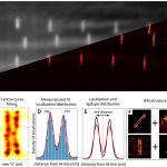
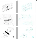
Object detection is an image analysis task with a wide range of applications, which is difficult to accomplish with traditional programming. Recent breakthroughs in machine learning have made significant progress in this area. However, these algorithms are generally compatible with traditional pixelated images and cannot be directly applied for pointillist datasets generated by single molecule localization microscopy (SMLM) methods. Here, we have improved the averaging method developed for the analysis of SMLM images of sarcomere structures based on a machine learning object detection algorithm. The ordered structure of sarcomeres allows us to determine the location of the proteins more accurately by superimposing SMLM images of identically assembled proteins. However, the area segmentation process required for averaging can be extremely time-consuming and tedious. In this work, we have automated this process. The developed algorithm not only finds the regions of interest, but also classifies the localizations and identifies the true positive ones. For training, we used simulations to generate large amounts of labelled data. After tuning the neural network’s internal parameters, it could find the localizations associated with the structures we were looking for with high accuracy. We validated our results by comparing them with previous manual evaluations. It has also been proven that the simulations can generate data of sufficient quality for training. Our method is suitable for the identification of other types of structures in SMLM data.