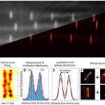
Localization based superresolution technique provides the highest spatial resolution in optical microscopy. The final image is formed by the precise localization of individual fluorescent dyes, therefore the quantification of the collected data requires special protocols, algorithms and validation processes. The effects of labelling density and structured background on the final image quality were studied theoretically using the TestSTORM simulator. It was shown that system parameters affect the morphology of the final reconstructed image in different ways and the accuracy of the imaging can be determined. Although theoretical studies help in the optimization procedure, the quantification of experimental data raises additional issues, since the ground truth data is unknown. Localization precision, linker length, sample drift and labelling density are the major factors that make quantitative data analysis difficult. Two examples (geometrical evaluation of sarcomere structures and counting the γH2AX molecules in DNA damage induced repair foci) have been presented to demonstrate the efficiency of quantitative evaluation experimentally.
Novák, T., Varga, D., Bíró, P., H. Kovács, B. B., Majoros, H., Pankotai, T., … & Erdélyi, M. (2022). Quantitative dSTORM superresolution microscopy. Resolution and Discovery. https://doi.org/10.1556/2051.2022.00093


