The quantitative detection of radiation caused DNA double-strand breaks (DSB) by immunostained γ-H2AX foci using direct stochastic optical reconstruction microscopy (dSTORM) provides a deeper insight into the DNA repair process at nanoscale in a time-dependent manner.
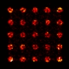
Sarcomeres are extremely highly ordered macromolecular assemblies where structural organization is intimately linked to their functionality as contractile units. Although the structural basis of actin and Myosin interaction is revealed at a quasiatomic resolution, much less is known about the molecular organization of the I-band and H-zone. We report the development of a powerful nanoscopic approach, combined with a structure-averaging algorithm, that allowed us to determine the position of 27 sarcomeric proteins in Drosophila melanogaster flight muscles with a quasimolecular, ∼5- to 10-nm localization precision. With this protein localization atlas and template-based protein structure modeling, we have assembled refined I-band and H-zone models with unparalleled scope and resolution. In addition, we found that actin regulatory proteins of the H-zone are organized into two distinct layers, suggesting that the major place of thin filament assembly is an M-line–centered narrow domain where short actin oligomers can form and subsequently anneal to the pointed end.
Nanoscopy reveals the layered organization of the sarcomeric H-zone and I-band complexes – Szilárd Szikora, Tamás Gajdos, Tibor Novák, Dávid Farkas, István Földi, Peter Lenart, Miklós Erdélyi, József Mihály
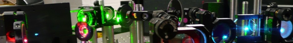
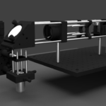
Super-resolution localization microscopy provides a powerful tool to study biochemical mechanisms at single molecule level. Although the lateral position of the fluorescent dye molecules can be determined routinely with high precision, measurement of other modalities such as 3D and multicolor without the degradation of the original super-resolved image is still in the focus. In this paper a dual-objective multimodal single molecule localization microscopy (SMLM) technique has been developed, optimized and tested. The proposed optical arrangement can be implemented onto a conventional inverted microscope without serious system modification. Read more
May 31, 2015Tamás GajdospublicationComments Off on Origin and compensation of imaging artefacts in localization-based super-resolution microscopy
Interpretation of high resolution images provided by localization-based microscopy techniques is a challenge due to imaging artefacts that can be categorized by their origin. They can be introduced by the optical system, by the studied sample or by the applied algorithms. Some artefacts can be eliminated via precise calibration procedures, others can be reduced only below a certain value. Images studied both theoretically and experimentally are qualified either by pattern specific metrics or by a more general metric based on fluorescence correlation spectroscopy.
The reduction of out of focus signal is a general task in fluorescence microscopy and is especially important in the recently developed super-resolution techniques because of the degradation of the final image. Several illumination methods have been developed to provide decreased out of focus signal level relative to the common epifluorescent illumination. In this paper we examine the highly inclined and the total internal reflection illumination techniques using the ray tracing method. Two merit functions were introduced for the quantitative description of the excitation of the selected region. We studied the feasibility of illumination methods, and the required corrections arising from the imperfections of the optical elements.
Localization-based super-resolution microscopy image quality depends on several factors such as dye choice and labeling strategy, microscope quality and user-defined parameters such as frame rate and number as well as the image processing algorithm. Experimental optimization of these parameters can be time-consuming and expensive so we present TestSTORM, a simulator that can be used to optimize these steps. TestSTORM users can select from among four different structures with specific patterns, dye and acquisition parameters. Example results are shown and the results of the vesicle pattern are compared with experimental data. Moreover, image stacks can be generated for further evaluation using localization algorithms, offering a tool for further software developments.
Localization microscopy software generally contains three elements: a localization algorithm to determine fluorophore positions on a specimen, a quality control method to exclude imprecise localizations, and a visualization technique to reconstruct an image of the specimen. Such algorithms may be designed for either sparse or partially overlapping (dense) fluorescence image data, and making a suitable choice of software depends on whether an experiment calls for simplicity and resolution (favouring sparse methods), or for rapid data acquisition and time resolution (requiring dense methods). We discuss the factors involved in this choice. We provide a full set of MATLAB routines as a guide to localization image processing, and demonstrate the usefulness of image simulations as a guide to the potential artefacts that can arise when processing.
Localization based super-resolution microscopy techniques require precise drift correction methods because the achieved spatial resolution is close to both the mechanical and optical performance limits of modern light microscopes. Multi-color imaging methods require corrections in addition to those dealing with drift due to the static, but spatially dependent, chromatic offset between images. We present computer simulations to quantify this effect, which is primarily caused by the high- NA objectives used in super-resolution microscopy. Although the chromatic offset in well corrected systems is only a fraction of an optical wavelength in magnitude (<50 nm) and thus negligible in traditional diffraction limited imaging, we show that object colocalization by multi-color super-resolution methods is impossible without appropriate image correction. The simulated data are in excellent agreement with experiments using fluorescent beads excited and localized at multiple wavelengths. Finally we present a rigorous and practical calibration protocol to correct for chromatic optical offset, and demonstrate its efficacy for the imaging of transferrin receptor protein colocalization in HeLa cells using two-color direct stochastic optical reconstruction microscopy (dSTORM). more…
Privacy & Cookies: This site uses cookies. By continuing to use this website, you agree to their use.
To find out more, including how to control cookies, see here:
Cookie Policy